
The over-arching goal of the lab is to understand how tissues and organs are sculpted in the developing embryo through an integration of genetic, molecular, and biophysical cues regulating complex cell behaviors. Using live in vivo imaging, gene misexpression, and a combination of experimental and computational mechanics, our lab aims to understand how forces that shape the embryo are specified by developmental signals, and how these forces in turn feedback on cell behaviors underlying growth, morphogenesis, and cell fate determination. Combining a classical model of embryology, the chick embryo, with state of the art approaches in imaging, engineering, and multi-omics, we focus our efforts on several distinct developmental events, exploiting practical advantages of each to investigate complementary conceptual questions that are fundamental to embryonic development.
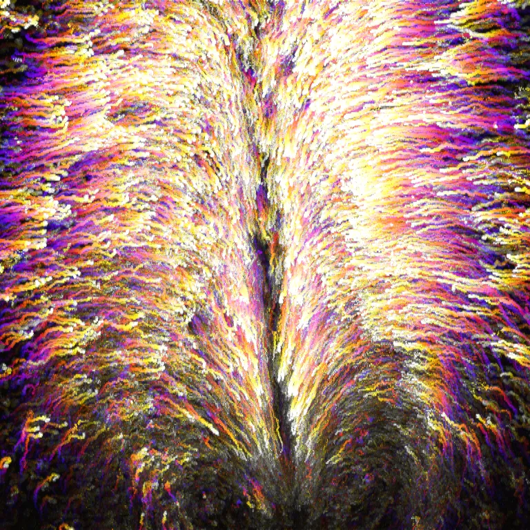
Morphogenesis of the gut tube
The vital functions of breathing, digestion, and nutrient absorption are carried out by the coordinated activity of several organs that, despite their functional and morphological diversity, arise from a single embryonic structure, the gut tube. Despite extensive work on the initial establishment of the endoderm during gastrulation and later organogenesis of endoderm-derived organs, the intervening stages of gut tube formation are remarkably understudied, despite the fundamental importance of this process for establishing the body plan. Because the endoderm lies on the ventral surface of amniote embryos at the onset of tube formation, and the early chick embryo, like humans and other non-rodent mammals, is flat, the endoderm is amenable to live in vivo imaging. As a result, one can directly visualize tissue deformations and underlying cell behaviors to ask how chemo-mechanical coupling enables collective cell migration, intercalation, EMT, and other conserved cell behaviors that coordinate the formation of the foregut, midgut, and hindgut.
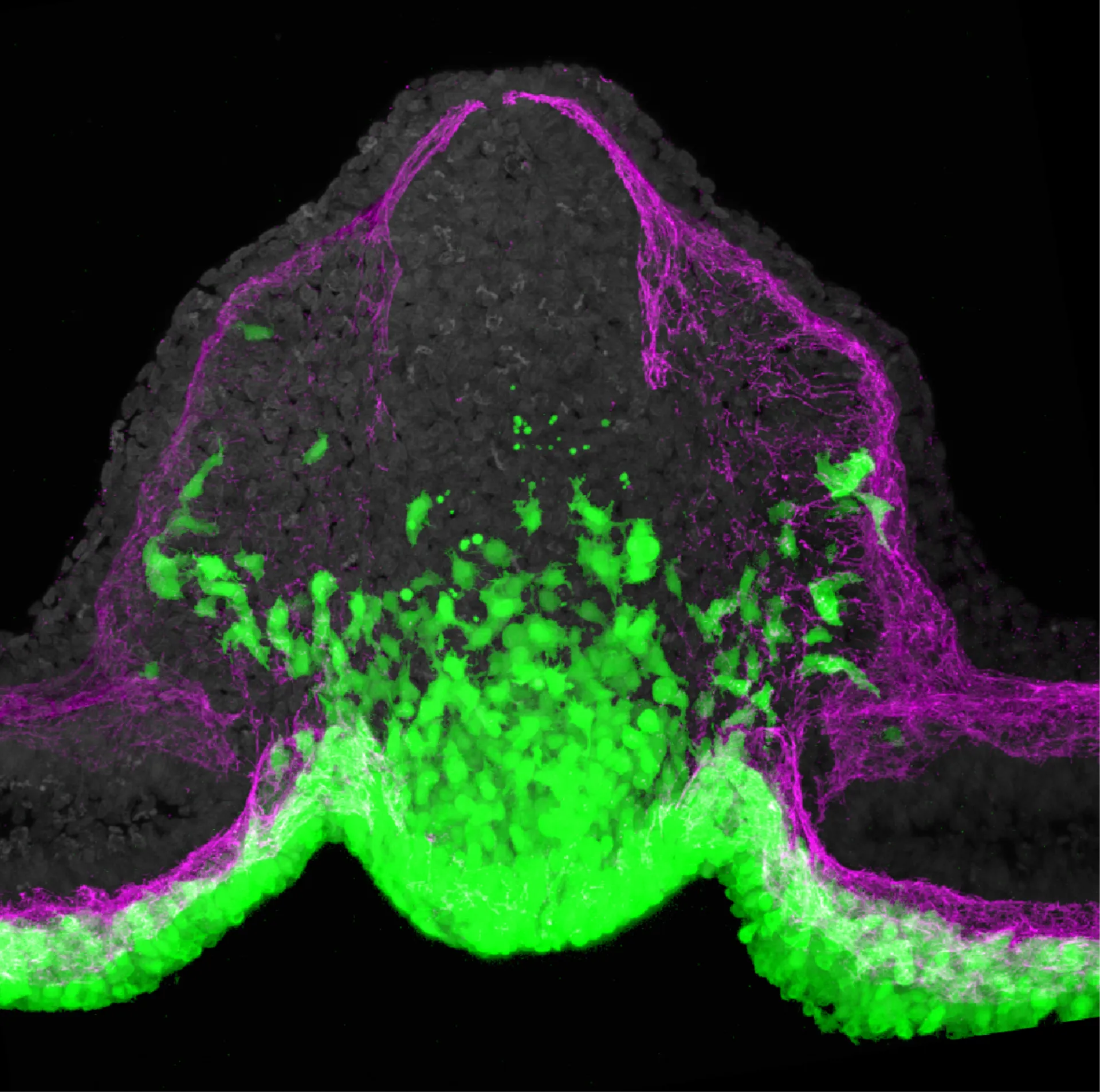
The node endoderm: a new stem cell population
In the stages spanning between gastrulation and organogenesis of definitive digestive organs, the endoderm has been so understudied that it remains full of mystery! For example, we recently discovered a new cell population within the endoderm that does some very surprising things: it undergoes a massive and previously unappreciated epithelial to mesenchymal transition (EMT), invading the neighboring mesoderm, it sheds it's endodermal identity as it does so, it gives rise to a remarkably broad range of tissue types, including traditionally mesodermal and ectodermal tissues, and it plays an essential role in elongation of the primary body axis, a process traditionally attributed to mesoderm. Combining live imaging, single cell RNA sequencing, and lineage barcoding together with classical embryology approaches, we are using this new progenitor population to understand a range of basic cell behaviors fundamental to both development and cancer progression, including EMT, cell fate determination and plasticity, directed cell migration, and fate convergence across surprisingly distant progenitor populations.
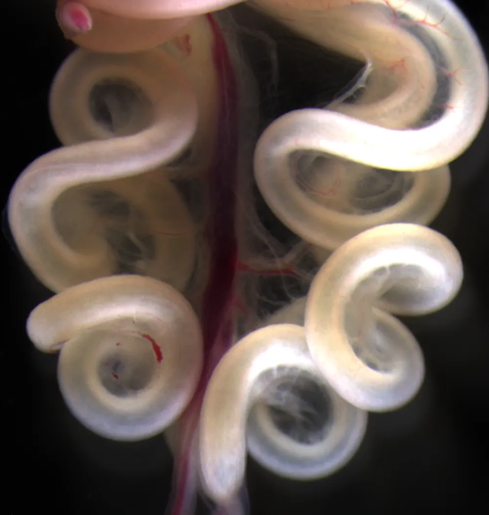
Organogenesis of the small intestine
Buckling is a core physical mechanism shaping a range of tissues and organs during embryogenesis, from folds on the cortical surface of the brain to branching of the airways. While in many cases, the physics of buckling morphogenesis have been well described, the integration of mechanics with underlying biology, an inherently cross-disciplinary challenge, is relatively limited. As a result, our understanding of biological controls on the mechanics of buckling remains limited. We use one tractable example of buckling morphogenesis, the looping of the small intestine, as a model to understand how mechanics are encoded, regulated, and enacted at a biological level to shape tissues. In this system, we can experimentally measure the geometric, mechanical and growth properties necessary to fully define the physics of buckling. Combined with embryology and gene misexpression approaches, we can then quantitatively investigate the molecular cues that control buckling, the determinants of complex material properties in embryonic tissues, how physical forces feedback on biological controls, the role of extracellular matrix in morphogenesis, and how evolution of morphological diversity across species arises through changes to biophysical phenomena.
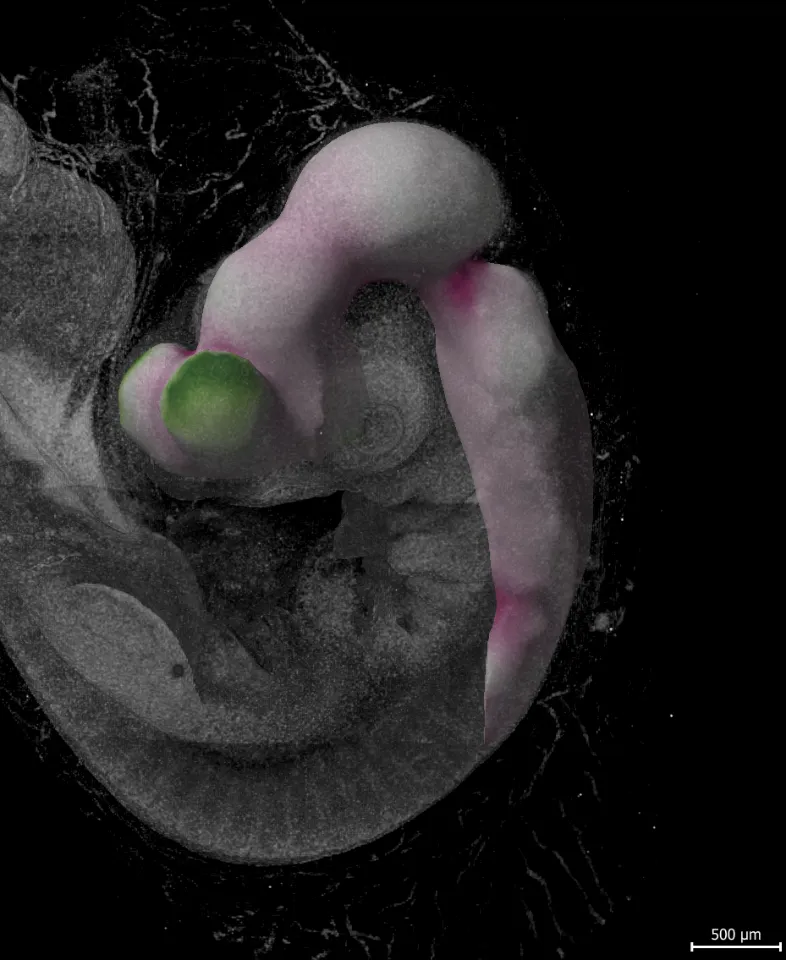
Mechanobiology of embryonic brain development
Despite the profound complexity of the central nervous system, its embryological origin is seemingly simple: a cylindrical tube inflated internally by cerebrospinal fluid (a "thick-walled pressure vessel" in engineering terms). This project aims to investigate how neuroepithelial progenitors in the cranial neural tube integrate forces from hydrostatic pressure with diffusible signaling factors to control growth, morphogenesis, and differentiation as the basic morphological plan of the brain is first established during development.